正文
PC14HM
更具
EMT
的特点,之后我们将使用来源于这两种细胞系的外泌体作用于非恶性细胞)
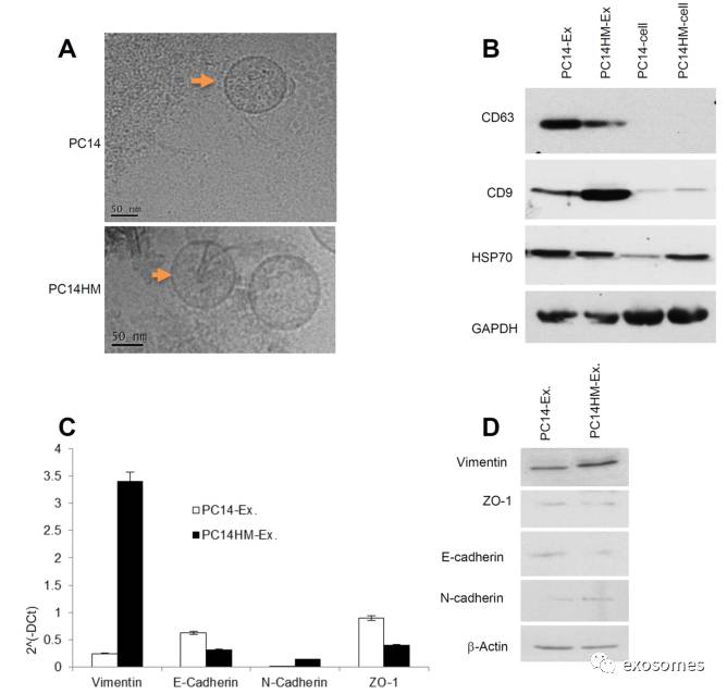
Figure2: Characterization of exosomes derived from PC14 and PC14HM cells
. A. Cryo-Transmission Electron Microscopy (cryo-TEM). TEM images of exosomes derived from PC14 and PC14HM cells. B. Western Blot analysis for exosomes marker in exosomes and cell lysates from PC14 and PC14HM cells. Twenty micrograms of total protein from exosomes or cell lysate were analyzed byWestern Blot using different exosome markers. GAPDH was used as an internal loading control. C. The relative mRNA expression of epithelial (E-cadherin, microgramsofZO-1), and mesenchymal (N-cadherin, Vimentin) markers by qRT-PCR in exosomalRNA isolated from PC14 and PC14HM cells. Normalization with housekeeping gene(GAPDH). The bars represent as mean ± SD of experiment performed in triplicate.D. Western Blot analysis of EMT marker in exosomal protemicrograms of total protein associated with exosomes were analyzed by Western Blot.β-Actin was used as an internal loading control. Ex indicates exosomes.
(
通过冰冻电镜检测收集到的
exosomes,
通过
western blot
检测
exosomes
标志物,通过
qRT-PCR
检测上皮或间质标志物,并进一步通过
western blot
检测标志物的表达量,从而分析两种细胞系的差异特点
)
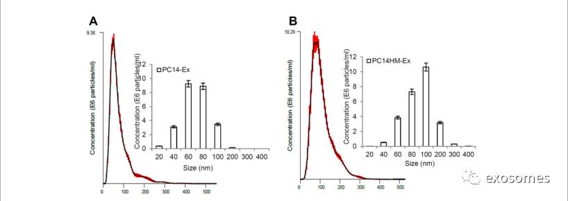
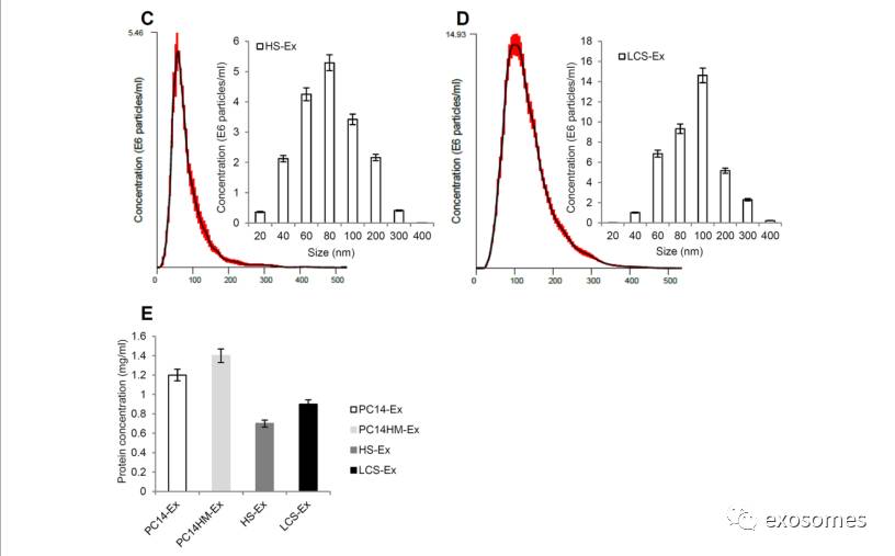
Figure 3: Exosome characterization by nanoparticletracking analysis.
Barchart showing the average percentage of nanoparticles within 20–300 nm size inin vitro exosome preparation. Concentration and size distribution of exosomesderived from A. PC14, B. PC14HM, C. healthy human serum, (HS), and D. lungcancer serum (LCS) were measured by nanoparticle tracking analysis (NTA).Exosomal concentration showed a peak at 60 +/− 0.5 nm (PC14 cell derivedexosomes, PC14-Ex), 100 +/−0.2 nm (PC14HM cell derived exosomes, PC14HM-Ex), 80+/−0.3 nm (healthy serum derived exosomes, HS-Ex) and 100 +/−0.7 nm (lungcancer serum derived exosomes, LCS-Ex). Bar Chart showing the particlenumber/ml of PC14, PC14HM, HS and LCS derived exosomes. E. ProteinConcentration of exosomes derived from PC14, PC14HM, healthy serum (HS) andlung cancer serum (LCS). Values are mean ± SD, all values are representative ofat least three independent experiments with four replicates.
(通过
NanoSight tracking analysis (NTA)
进一步对从
4
组细胞中提取出来的
exosomes
的大小及含量进行检测,并通过
BCA
检测
exosomes
内的蛋白含量)