正文
版主微信号:
fsslong2
【临床病史】
患者,42岁女性,有头痛史,有头痛史和人格改变,行CT扫描。42-year-old female with history of headache and personality change undergoes a CT brain scan.
【影像图片】
MRI图像

【影像表现】
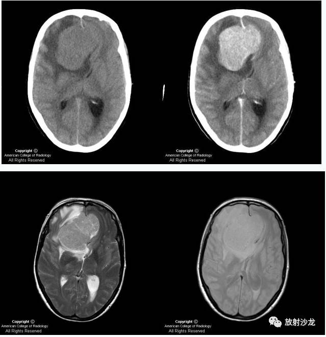
Axial non contrast enhanced CT [Figure 1] shows a large lesion isointense to the brain in the frontal lobes crossing the midline.
Contrast enhanced axial image Figure 2] shows avid contrast enhancement of the lesion.
T2W axial image (Figure 3) shows a mixed signal large lesion with little surrounding edema.
Proton density axial image Figure 4) shows a lesion which is largely isointense to the surrounding brain parenchyma.
The lesion (Figure 5 and Figure 6) is low signal on TIW image with marked contrast enhancement.
Post contrast TIW sagittal image (Figure 7) shows the lesion to be extra axial with contrast enhancement


