正文
结果:
1.Characteristics of extracellular vesiclesfrom control and cisplatin-treated cells.
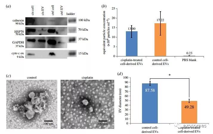
Figure 1. Characterizationof cisplatin and control EVs derived from A2780 cells. (a) Control andcisplatin-treated cell and EV protein lysates were characterized by Westernblotting; samples were probed for GAPDH, calnexin, HSP70 and cytochrome coxidase. (b) Quantification of EVs secreted by control and cisplatin-treated A2780cells by nanoparticle tracking analysis (at least two replicates). (c) Imagesof electron microscopy grids of control and cisplatin EVs visualized by transmissionelectron microscopy. (d) Average diameter of EVs secreted by cisplatin-treatedand control A2780 cells measured on electron microscopy grids (c). (Onlineversion in colour.)
2.
Extracellular vesicles released by cisplatin-treated cells have thecapacity to induce invasion.
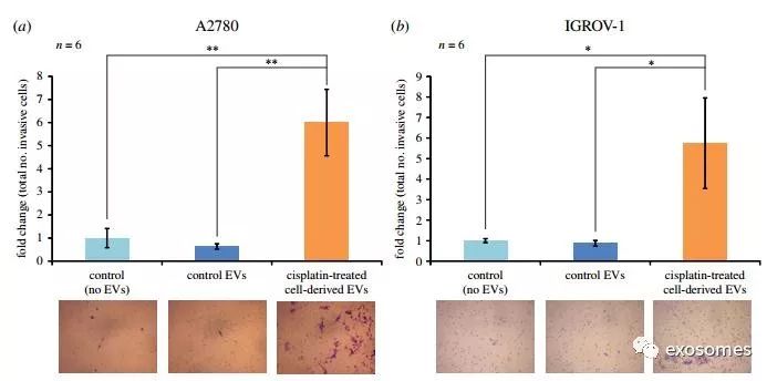
Figure 2. The effect ofcisplatin-treated cell-derived EVs upon the invasive capacity of ovarian cancercell lines. The Matrigel transwell invasion assay was used to determine theeffect of cisplatin-treated cell-derived EVs on invasive potential of twoovarian cancer cell lines, A2780 (a) and IGROV-1 (b). Extracted EVs wereadministered to approximately 1 million cells and after 24 h, 100 000 cellswere distributed into each insert of the transwell assay and another dose ofEVs was added. After 24 h, the Matrigel membranes were cleared of noninvasivecells and invasive cells were stained with crystal violet. The number ofinvasive cells on each membrane was counted. The graphs represent fold changein terms of the total number of cells that invaded the Matrigel membranefollowing treatment with either control or cisplatin-treated cell-derived EVs.Each sample group contained six biological replicates. Error bars representstandard error of the mean of the biological replicates. p-Values werecalculated using Student’s t-test. Representative images are shown below eachgroup. (Online version in colour.)


